Executable Ancestor
The complete brain of a fruit fly, all 166,700 neurons, simulated on a GPU and force-fed a film. It has no body, so all it can do is respond.
- Role
- Solo project: concept, direction, development, visuals
- Skills
- Core Dev + Events
- Year
- 2026, in progress
- Input
- A film, processed offline into a brain recording
- Data
- MaleCNS fruit fly connectome
- Tech stack
- TouchDesigner, neuPrint connectome API, NVIDIA GPU, Python

The project
What does it mean to run a nervous system older than language on a machine? Executable Ancestor takes the complete connectome of a male fruit fly, 166,700 neurons joined by 25.6 million real synapses, and runs it on a GPU as simplified neurons. The brain is given no body. The run is recorded, then replayed as a ghost built from the neurons’ own shapes.
It started with a discovery: a public demo from boat.horse that ran the complete MaleCNS connectome in a closed loop. Its code was not released, so the work was built on DOOMFLY, an open-source (MIT) connectome runner by nftechie, whose neuron kernel and graph loader the simulation still uses.
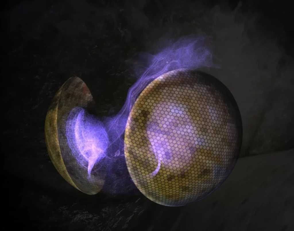
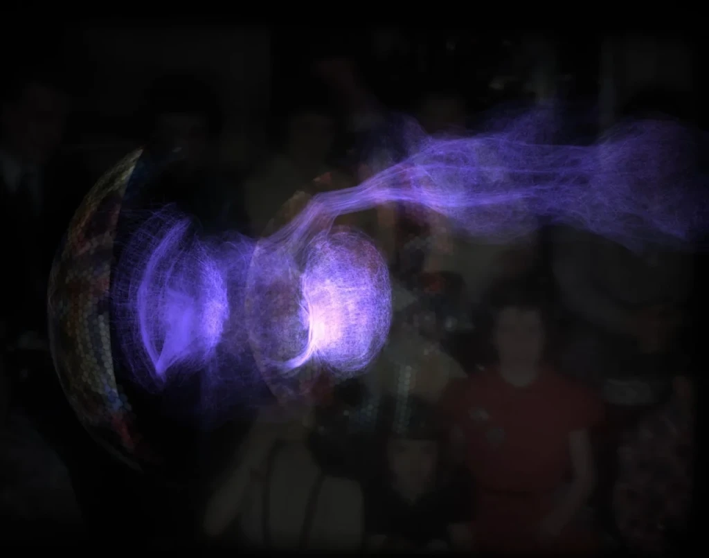
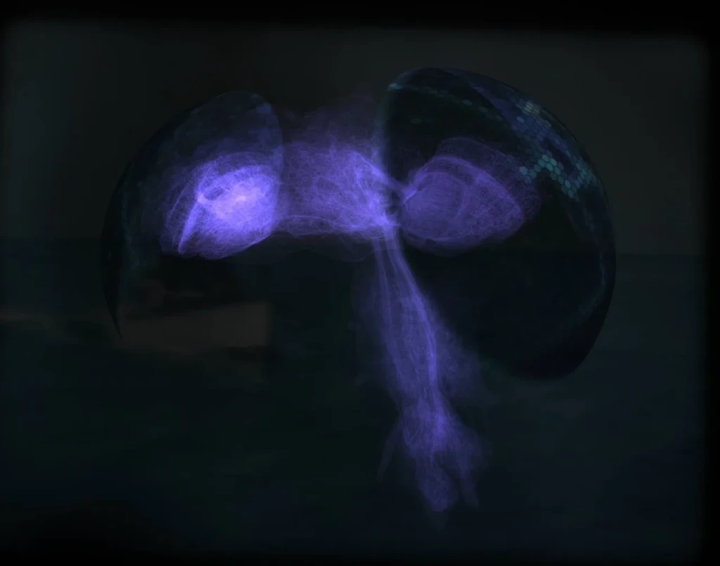
The work has three stages: a simulation of the wiring in a toy world, a portrait of the recorded activity, and a newer chapter that places the brain between two compound-eye domes showing a film. The film becomes input to the optic nerves, and the nerves light the film back.

The whole nervous system is a small part of the body: the brain sits in the head and the nerve cord runs back from the neck into the thorax. Neuron counts are for the female BANC dataset.
Image: Bates et al., “Distributed control circuits across a brain-and-cord connectome”, Nature (2026), Fig. 1f, CC BY 4.0. Cropped and recolored for a dark background.
The wiring
The data is the MaleCNS connectome, published in 2026 by HHMI Janelia, the University of Cambridge, the MRC Laboratory of Molecular Biology and Google Research. It maps every neuron in a male fruit fly’s central nervous system and the synapses between them.
Skeletons and meshes are public, and cell types and positions are available through the neuPrint query API. Of the 166,700 neurons, 139,662 have a known cell-body position. The first portrait draws those cell bodies, joined by about 24,000 of the strongest of the 25.6 million real connections.
Color carries information. Each neuron is tinted by the region it belongs to, and a connection takes the tint of the neurons it joins.
- Optic lobes
- Central brain
- Descending
- Ascending
- Nerve cord
- Firing


The neurons
Each neuron is modeled as a leaky integrate-and-fire unit. It adds up the signals arriving through its synapses, leaks charge as time passes, and fires when it crosses a threshold. Excitatory synapses push it toward firing, and inhibitory ones, about 38% of the total, push it away from it.
Parameters follow Shiu et al. (Nature, 2024), and a single GPU steps the whole network at roughly real time. Integrate-and-fire is a crude description of a real neuron, and no claim of biophysical accuracy is made.

The ghost
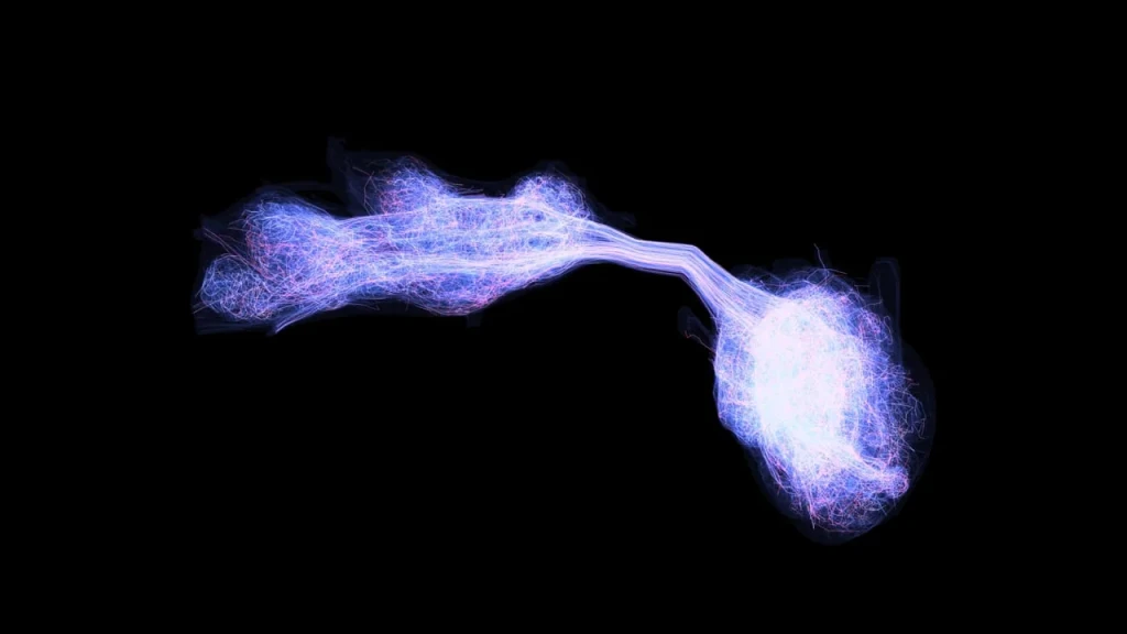
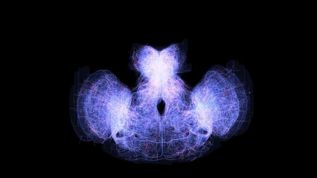
Rather than giving the brain a body, the portrait draws it as a ghost. 19,372 neurons appear as their real skeletons, simplified from the public data, inside glassy shells of the regions they occupy. Fourteen hero neurons from the looming pathway are drawn as full surface meshes.
A drone camera travels through the structure. The pulses running along the axons are a display layer over recorded spikes, timed by distance from the cell body, and not a measured signal path.


Left: the first render with real neuron shapes. Right: pulses traveling along them. The brain is on the left of each frame and the nerve cord, the fly’s counterpart of a spinal cord, runs off to the right. The cord is drawn from real skeletons, but nothing in the simulation reads from it.
133x slower
The simulation steps in half-millisecond slices. Played back at 15 slices per second, the brain runs about 133 times slower than the simulation did, slow enough for individual pulses to be followed by eye. The result is a replay of a recording, not a live run.
The film chapter makes a different trade. A film slowed 133 times would be destroyed, so the film plays at normal speed and the brain’s activity is replayed about 8 times slower than simulated. From the brain’s point of view, the film plays about 8 times too fast. This is an artistic decision, not a physiological one.

The eyes
The idea is to force the brain to watch a film and observe how it responds. The fly’s own wiring decides which neurons react to what the film shows, and in roughly what order.
Two hex-faceted domes, a stylized version of the fly’s compound eyes, wrap the brain. Their center, axis and size are fitted to 1,773 medulla columns in the connectome, so the eyes sit where the real visual system places them.
Only the forward tilt, 25°, is a judgment, taken from a photograph of a fly. The connectome’s own axes suggest closer to 50°.


Fitting the domes to the anatomy, from above, front and side. The orange and blue dots are the 1,773 real medulla columns, one set per eye. Red is the first guess. Green is a fit covering all the optic lobes and yellow a fit to the lamina and medulla only.
To make that possible, each film is reduced to a simple measure of movement. Frame by frame, the difference between consecutive images is taken after film grain, flicker and vignetting are filtered out. The result is a motion map: only what changes between frames is visible, tinted with the color of the original film.
The domes display that map. It is stretched across both eyes, one half of the frame for each, with a small overlap down the middle. Each hexagon samples the map once, at its own center, so each one carries a single flat tone: its fraction of the picture, in the color of the footage beneath it. A hex over a moving boat glows, and a hex over still water goes dark. Together the facets break the frame into a mosaic, an artistic echo of a compound eye. The background keeps the original film.
The same signal drives the simulation. It enters as injected current: motion is averaged over the left and right halves of the picture, and each half drives T4 and T5, the neurons that detect movement, in the matching eye. A sudden rise in motion, the short-term average climbing above the long-term one, is sent to LC4 and LPLC2, the neurons tuned to looming objects, which is how an approaching shape would register. A small steady inhibitory current lets activity fall again when the picture calms. The nerves behind each eye then light up in response, in the color of the hexes above them.
This is a crude detector. Brain activity follows how much of the picture is moving, not whether something is approaching: a control clip of a swinging crane bucket drove the looming neurons at least as strongly as a motorboat heading toward the camera. Only a left and right split reaches the simulation, with no spatial detail, and gains are scaled per clip so that quiet films still register.
Despite this being a crude detector, the haunting visuals it produces are the payoff. Light moves through the real wiring of a fly’s brain as the film moves, flaring where a boat crosses the frame and fading as the picture settles. 42 clips have been processed this way, each recorded to its own brain recording.

Gallery
Ending Notes
- The looming pathway works. With looming input on, the simulated fly never touched a wall in a test arena; with it removed, it hit walls 354 times, across two random seeds. The adapter that turns spikes into movement shapes this result.
- The optic lobes fire in lockstep volleys at about 435 Hz. Constant, very large drive into identical, noise-free neurons locks them to their refractory limit. This is an artifact of the model, not a finding about the fly.
- The network never goes quiet once driven. About 15,000 neurons keep firing with no input, a sign of runaway recurrent excitation in the model.
- Looming detection from film is crude. Brain activity follows how much of the picture moves, and a control clip of a swinging crane bucket drove the looming neurons at least as strongly as an approaching boat.
The wiring, and the position and shape of each neuron, belong to the dataset. The neuron model is simplified, the stimulus is synthetic, and the timing, color and camera are artistic choices. The result is an artwork built from the data, not a validated model of a fly.
The software was developed with AI coding tools, under the artist’s direction.
Neuron data: MaleCNS connectome, FlyEM at HHMI Janelia Research Campus, University of Cambridge (Zoology), MRC Laboratory of Molecular Biology and Google Research (CC BY 4.0). Weights, mappings and visuals are the artist’s own transformations and are not endorsed by the dataset creators. Neuron model after Shiu et al., Nature 2024. Simulation built on DOOMFLY (nftechie, MIT License). Footage: Tom Kirsch; family film by John Kirsch (c. 1940s). Made in TouchDesigner. Concept, direction and visuals: Tom Kirsch.